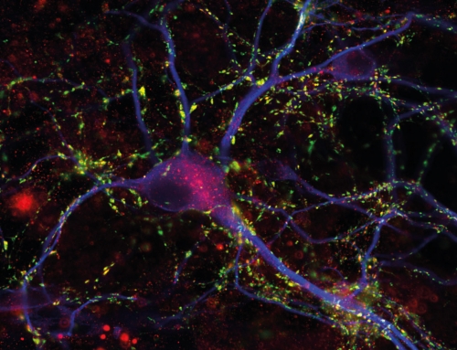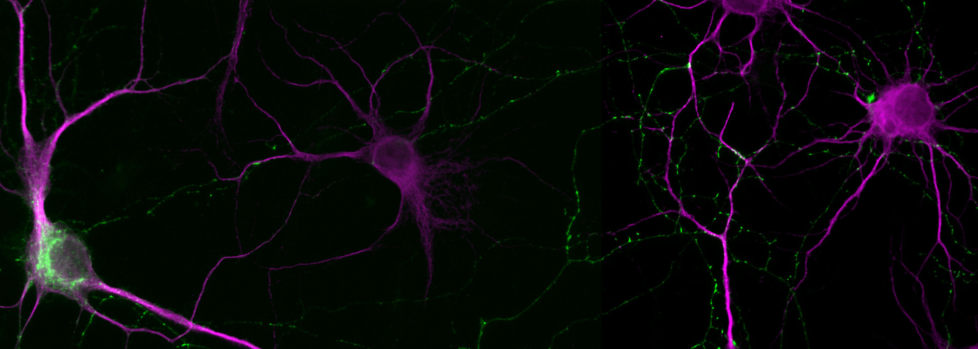
Presynaptic Targeting of GFP-VAMP in a Transfected Neuron
The experiment shows the distribution of a recombinant version of a synaptic vesicle protein, i.e. GFP-tagged VAMP/Synaptobrevin (green), in a transfected neuron (left). All neurons are stained using MAP2 (magenta), to show their dendrites and soma. GFP-VAMP puncta observed in the right part of the field of view represent synaptic vesicle accumulations in the axon of the transfected neuron.
Image details
Courtesy Donatus Riemann and Thomas Dresbach / UMG, CNMPB