Labeling neuronal compartments
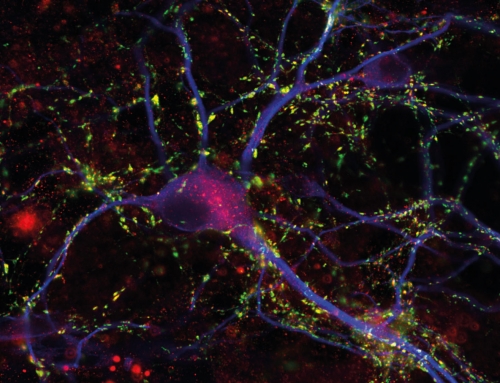
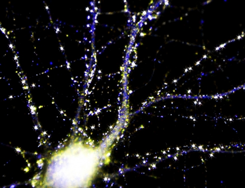
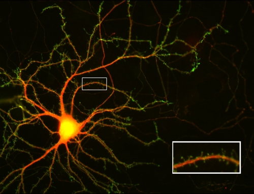
Triple fluorescence of a neuronal culture transfected with GFP (green) using the calcium- phosphate method. GFP was used to fill and detect all processes of the transfected neuron at the bottom of the image. MAP2 (red) labels dendrites, the soma, and a short, proximal part of the axon of all neurons. TRIM46 (blue) selectively labels proximal parts of the axon of all neurons.
Image details
Courtesy Donatus Riemann and Thomas Dresbach / UMG, CNMPB